Diagnosis
During
the physical exam, your doctor may ask you to move in certain ways to
check for pain and evaluate your range of motion (active range of
motion). Your doctor might then ask you to relax your muscles while he
or she moves your arm (passive range of motion). Frozen shoulder affects
both active and passive range of motion.
In
some cases, your doctor might inject your shoulder with a numbing
medicine (anesthetic) to determine your passive and active range of
motion.
Frozen
shoulder can usually be diagnosed from signs and symptoms alone. But
your doctor may suggest imaging tests — such as X-rays or an MRI — to
rule out other problems.
Treatment
-
Shoulder exercises
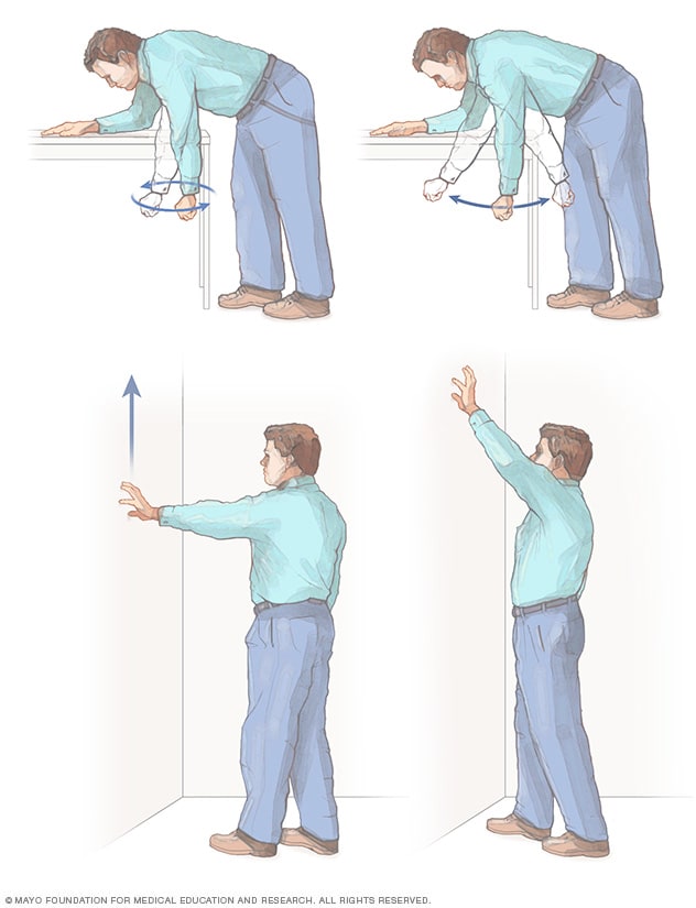
Most
frozen shoulder treatment involves controlling shoulder pain and
preserving as much range of motion in the shoulder as possible.
Medications
Over-the-counter
pain relievers, such as aspirin and ibuprofen (Advil, Motrin IB,
others), can help reduce pain and inflammation associated with frozen
shoulder. In some cases, your doctor may prescribe stronger
pain-relieving and anti-inflammatory drugs.
Therapy
A
physical therapist can teach you range-of-motion exercises to help
recover as much mobility in your shoulder as possible. Your commitment
to doing these exercises is important to optimize recovery of your
mobility.
Surgical and other procedures
Most frozen shoulders get better on their own within 12 to 18 months. For persistent symptoms, your doctor may suggest:
- Steroid injections. Injecting corticosteroids into your shoulder joint may help decrease pain and improve shoulder mobility, especially in the early stages of the process.
- Joint distension. Injecting sterile water into the joint capsule can help stretch the tissue and make it easier to move the joint.
- Shoulder manipulation. In this procedure, you receive a general anesthetic, so you'll be unconscious and feel no pain. Then the doctor moves your shoulder joint in different directions, to help loosen the tightened tissue.
- Surgery. Surgery for frozen shoulder is rare, but if nothing else has helped, your doctor may recommend surgery to remove scar tissue and adhesions from inside your shoulder joint. Doctors usually perform this surgery with lighted, tubular instruments inserted through small incisions around your joint (arthroscopically).
Lifestyle and home remedies
Continue
to use the involved shoulder and extremity as much as possible given
your pain and range-of-motion limits. Applying heat or cold to your
shoulder can help relieve pain.
Alternative medicine
Acupuncture
Acupuncture
involves inserting extremely fine needles in your skin at specific
points on your body. Typically, the needles remain in place for 15 to 40
minutes. During that time they may be moved or manipulated. Because the
needles are hair thin and flexible and are generally inserted
superficially, most acupuncture treatments are relatively painless.
Transcutaneous electrical nerve stimulation (TENS)
A
TENS unit delivers a tiny electrical current to key points on a nerve
pathway. The current, delivered through electrodes taped to your skin,
isn't painful or harmful. It's not known exactly how TENS works, but
it's thought that it might stimulate the release of pain-inhibiting
molecules (endorphins) or block pain fibers that carry pain impulses.
Preparing for your appointment
While you might first consult your family physician, he or she may refer you to a doctor who specializes in orthopedic medicine.
What you can do
Before your appointment, you may want to write down:
- Detailed descriptions of your symptoms
- Information about medical problems you've had
- Information about the medical problems of your parents or siblings
- All the medications and dietary supplements you take
- Questions to ask the doctor
What to expect from your doctor
Your doctor may ask some of the following questions:
- When did your symptoms begin?
- Are there activities that worsen your symptoms?
- Have you ever injured that shoulder? If so, how?
- Do you have diabetes?
- Have you had any recent surgeries or periods of restricted shoulder motion?